我的博文
肌周皮细胞瘤
肌周皮细胞瘤 | |
概 述 | 肌周皮细胞瘤(myopericytoma)是一种位于皮下的良性肿瘤,由卵圆形至梭形的肌样细胞组成,瘤细胞常呈同心圆状围绕血管生长。肌周皮细胞瘤的概念由Granter等人于1998年提出,德国Friedrichshafen皮肤病理研究所的Kutzner于同年提出了血管周肌瘤(perivascular myoma)的概念,两者实质上为同一肿瘤。瘤细胞具有血管周肌样细胞(myoid cell)或肌周皮细胞分化的特征。肌周皮细胞瘤与血管平滑肌瘤、肌纤维瘤、球周皮细胞瘤、血管球瘤和所谓的婴幼儿型血管外皮瘤共同组成一个瘤谱。 |
临床表现 | 最常见于中年人。多发生于肢体皮下,表现为缓慢生长的无痛性结节。常为单个结节,多发者多为异时发生。Myopericytoma-like tumors, sometimes multiple, have been reported as arising in the setting of Epstein-Barr virus (EBV) infection. Such lesions likely represent morphologic variants of EBV-associated smooth muscle tumors. |
大体形态 | 结节周界清晰,直径在2cm以下,恶性者可达数厘米。 |
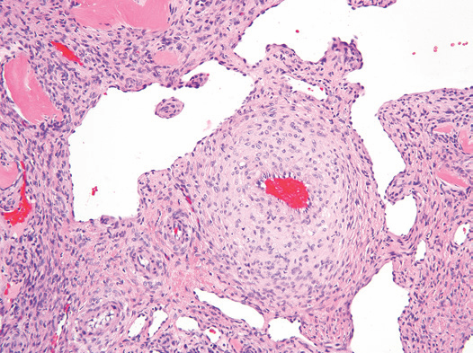
组织形态学 | 细胞形态:相对一致的卵圆形至梭形肌样细胞分布于大小不一的血管周围。肌样细胞胞质嗜伊红,核染色质均匀,核异型性不明显,核分裂象少见; 排列方式:经典形态至少在局部区域可见多层肌样细胞围绕小至中等大的血管呈同心圆状或漩涡状生长,同心圆或漩涡间的基质可伴有黏液样变性;部分区域瘤细胞丰富呈片状分布;局部区域血管呈分支状类似血管外皮瘤; 总的说来,本瘤在组织学上包括了肌周皮细胞瘤、婴幼儿型血管外皮瘤、肌纤维瘤和血管球瘤几种形态。 |

Figure Myopericytoma. The tumor is composed of short spindle cells with eosinophilic cytoplasm concentrically arranged around variably thick-walled or dilated, branching blood vessels.

Figure Myopericytoma. The tumor cells contain ovoid to elongated nuclei, pale cytoplasm, and ill-defined cell borders.

Fig. Myopericytoma. Muscle from walls of small-caliber vessels spin off into stroma of the lesion, giving it an appearance intermediate between hemangiopericytoma and angiomyoma.

Fig. Myopericytoma showing ectatic vessels surrounded by spindled cells.

Fig. Spindled cells within myopericytoma.
免疫组化 | 瘤细胞多为弥漫表达SMA、h-caldesmon,偶可灶性表达desmin和CD34。 不表达S100和CK。 |
分子遗传学 | 部分病例有t(7;12)(p21-22;q13-15)/ACTB-GLI1基因融合和BRAF突变。 SRF-RELA gene fusion in cellular myopericytoma, and PDGFRB mutations in so-called myopericytomatosis. The few classic cases examined in one study lacked PDGFRB mutations. |
治疗与预后 | 局部完整切除,境界不清或切除不干净可复发。核分裂象(良性多<1个/10HPF)易见可能提示恶性?应对患者进行长期随访。 |
参考文献
[1] 王坚,朱雄增. 软组织肿瘤病理学[M].2017.
[2] Hornick JL. Practical Soft Tissue Pathology: A Diagnostic Approach[M].2018.
[3] Enzinger and Weiss's Soft Tissue Tumors[M].2020.
[4] WHO Classification of Skin Tumours[M].2018.
我要评论


共0条评论