我的博文
2020年WHO分类软组织肿瘤病理学新进展(3)
Superficial CD34-positive Fibroblastic Tumor | 浅表性CD34阳性纤维母细胞肿瘤 |
This distinctive low-grade neoplasm of the skin and subcutis is rare, with few reported cases. | 这种独特的低度恶性皮肤和皮下肿瘤罕见,有少数病例报道。 |
Most have occurred in middle-aged adults, with a slight male predominance, and a predilection for the lower extremities, followed by upper extremities, buttock, and shoulder. | 大多数发生于中年人,男性略多见,好发于下肢,其次是上肢、臀部和肩部。 |
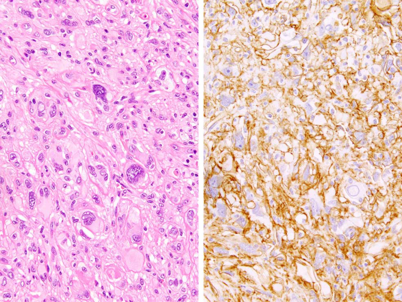
Histology demonstrates highly cellular fascicles and sheets of spindled to epithelioid cells with abundant eosinophilic, often glassy cytoplasm and moderate-to-marked pleomorphism (Fig. 5). | 组织学显示梭形到上皮样细胞呈束状和片状分布,细胞密度高,细胞质丰富嗜酸性,通常为玻璃状,中度到显著的多形性(图5)。 |
There is morphologic overlap reported with PRDM10-rearranged soft tissue tumors. | 形态学与PRDM10基因重排的软组织肿瘤有重叠。 |
By IHC, there is strong CD34 expression (Fig. 5B), and keratins are positive in 70% of cases. The prognosis in reported cases has been excellent. | IHC显示CD34强阳性表达(图5B),70%的病例表达角蛋白。文献报道该疾病预后良好。 |

FIGURE 5. Superficial CD34-positive fibroblastic tumor. A, The tumor is composed of sheets of variably pleomorphic and epithelioid cells with abundant, glassy eosinophilic cytoplasm. Mitotic activity is minimal, in contrast to undifferentiated pleomorphic sarcoma. B, IHC for CD34 is diffusely positive. Keratins are also often positive (not shown).
图5 浅表性CD34阳性纤维母细胞肿瘤。A、肿瘤由多少不等的多形性和上皮样细胞组成,呈片状分布,胞质丰富,呈玻璃状嗜酸性。与未分化的多形性肉瘤相比,核分裂象极少。B、IHC显示CD34呈弥漫阳性。角蛋白也常呈阳性(未显示)。
我要评论


共0条评论