我的博文
软组织透明细胞肉瘤
概述:
软组织透明细胞肉瘤(clear cell sarcoma of soft tissue)是一种具有色素细胞分化的软
组织肿瘤,由Enzinger于1965年首先报道。因其组织学和免疫表型相似于恶性黑色素瘤,以往
称为软组织恶性黑色素瘤。但在遗传学上,透明细胞肉瘤具有不同于恶性黑色素瘤的特异性染
色体易位t(12;22)(q13;q12)并产生EWSR1-ATF1融合性基因,故采用软组织透明细胞肉
瘤比软组织黑色素瘤更好。需要注意的是,发生于婴幼儿肾脏的透明细胞肉瘤虽然在名称上相
同,却是一种不同的肿瘤类型。
临床表现:
多发生于20~40岁青年人,女性较多见。好发于四肢末端,以足和踝常见。位置多较深,临床
上表现为缓慢生长的肿块,病程可达数年,可伴有疼痛。
大体形态:
肿瘤直径多为2~6cm,周界清晰,无包膜,分叶状或结节状,质实,常附着于腱鞘或腱膜,与
被覆皮肤不相连。切面灰白色,可有出血、坏死、囊性变。
组织形态学:
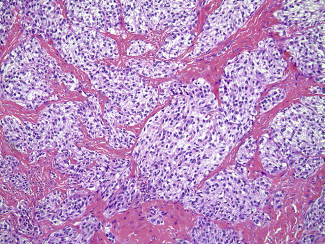
排列方式:瘤细胞呈束状、巢状或片状排列,其间为纤细或致密的纤维结缔组织间隔,网状纤
维染色能清晰显示巢状或器官样排列结构;少数病例由交织条束状排列的梭形细胞组成,可被
误诊为其他类型的梭形细胞肉瘤。
细胞形态:瘤细胞多边形、卵圆形或胖梭形,胞质常呈淡嗜伊红色,真正透亮者少见,核圆形
卵圆形,淡染或空泡状,可见明显的嗜伊红或嗜双色性核仁,核分裂象多<3~5个/10HPF。HE
染色时约20%病例于瘤细胞内可见黑色素颗粒。复发或转移性肿瘤中,瘤细胞可显示明显的异型性。1/3~1/2病例可见多核巨细胞,胞核位于胞质周边排列(花环状)。
间质:可有黏液样和玻璃样变性。

Fig.1 Fibrous tissue septa divide clear cell sarcoma into well-defined nests and groups of pale-staining tumor cells.

Fig.2 Clear cell sarcoma showing spindled cells with strikingly clear cytoplasm and tumor giant cells with peripherally placed nuclei of uniform size and shape.

Fig.3 Metastatic clear cell sarcoma with marked pleomorphism and essentially no spindling. Metastatic deposits of this type resemble carcinoma or melanoma.

图23-52 软组织透明细胞肉瘤,多数病例胞质呈淡嗜伊红色,可见明显核仁,部分区域瘤细胞可呈束状排列;
免疫组化:
瘤细胞弥漫表达S100和SOX10,还可表达HMB45、MelanA、MelCAM、MiTF、酪氨酸激酶、CD57
和NSE等。

Fig.4 Clear cell sarcoma with S-100 protein expression.
分子遗传学:
EWSR1-ATF1基因融合(90%以上病例);EWSR1-CREB1基因融合;
鉴别诊断:
①纤维肉瘤:无多核巨细胞,细胞内也无黑色素颗粒,不表达S100、HMB45;
②恶性周围性神经鞘膜瘤:肿块多与大神经相连或具有神经纤维瘤病表现。S100常为局灶阳性;不表达HMB-45和melan A.
③恶性黑色素瘤:与皮肤关系密切,被覆或邻近皮肤常有色素性或交界性病变存在。瘤细胞多
形性和异型性均较明显,核分裂象易见,瘤内常见较多的黑色素,而多核巨细胞相对少见。检测EWSR1基因有助于两者的鉴别诊断。
④Cellular blue nevus(细胞性蓝痣)can occur in a similar age and location and have certain common histologic features, including spindled cells and giant cells with clear cytoplasm. Cellular blue nevi typically are dermal-based lesions with a peripheral zone that resembles a neurofibroma by virtue of the interdigitation of slender, pigmented dendritic cells with surrounding collagen (Fig. 29.23). The cells lack atypia and have small, pinpoint nucleoli, in contrast to the macronucleoli of clear cell sarcoma (Fig. 29.24). Recurrent cellular blue nevi, however, can acquire more atypical cytologic features, so a distinction from clear cell sarcoma is not always possible. In these situations, review of the original material or molecular genetic analysis is essential.


Fig. 29.23 Cellular blue nevus showing partitioning of tumor by fibrous bands and prominent pigmentation (A,上图) but cells with less atypia and less prominent nucleoli (B,下图) than clear cell sarcoma.


Fig. 29.24 Comparison of cytologic features of clear cell sarcoma and a cellular blue nevus at the same magnification. A(上图), Clear cell sarcoma has prominent vesicular nuclei with a large single nucleolus. B(下图), Cellular blue nevus cells are smaller with a less vesicular nuclear chromatin pattern and small, pinpoint nucleoli.
⑤The recently described paraganglioma-like dermal melanocytic tumor(副神经节瘤样真皮色素细胞肿瘤), while having cells with a clear to eosinophilic cytoplasm, also has zellballen-like(细胞球样) nests of cells of distinctly low nuclear grade (Fig. 29.25). These lesions, based in the dermis, rarely extend to deep structures. At this point, all have behaved in a benign fashion.


Fig. 29.25 Paraganglioma-like dermal melanocytic tumor (A,上图) showing zellballen-like nests of slightly spindled to rounded cells (B,下图).
⑥Dermal perivascular epithelioid cell neoplasms(真皮血管周上皮样细胞肿瘤), typically show lesser degrees of nuclear atypia than do clear cell sarcoma, lack S-100 protein expression, and coexpress muscle and melanocytic markers.
⑦An unusual cutaneous melanocytic tumor resembling clear cell sarcoma, but
showing CRTC1-TRIM11 fusion, has recently been reported as well.
预后:
5年生存率为47%左右。复发病例、直径5cm以上或伴有坏死者预后不佳。
参考文献:
[1] 王坚,朱雄增. 软组织肿瘤病理学[M].2017.
[2] Enzinger and Weiss's Soft Tissue Tumors[M].2020.
我要评论


共0条评论