我的博文
病理医生也要关注“富营养化”现象
病理医生也要关注“富营养化”现象 | |
Eutrophication in histopathology | 组织病理学中的富营养化 |
Eutrophication is the term used to define the process by which some habitats such as lakes, reservoirs, streams or bays become overloaded with nutrient-rich water. | 富营养化是用来定义某些栖息地,如湖泊、水库、溪流或海湾,因富营养水而超载的过程。 |
360百科:富营养化是指湖泊、河流、水库等水体中氮磷等植物营养物质含量过多所引起的水质污染现象。由于水体中氮磷营养物质的富集,引起藻类及其他浮游生物的迅速繁殖,使水体溶解氧含量下降,造成藻类、浮游生物、植物、水生物和鱼类衰亡甚至绝迹的污染现象。 | |
This process can occur in both freshwater and saltwater habitats. When this occurs, large blooms of algae and aquatic plants grow as a result of an excess of nitrogen and phosphorus. Excess phosphorus often causes eutrophication in freshwater, whereas nitrogen is responsible for this phenomenon in saltwater. The sources of these excess nutrients in these habitats include agricultural runoff, overuse of synthetic fertilisers, and sewage leaks, among others. Additionally, certain climatic conditions such as high temperatures and low rain may also promote eutrophication. | 这一过程可以发生在淡水和咸水栖息地。当这种情况发生时,由于氮和磷的过量,藻类和水生植物大量繁殖。过量的磷常导致淡水富营养化,而氮则是造成这一现象的原因。这些栖息地中过量养分的来源包括农业径流、过度使用合成肥料、污水泄漏等。此外,某些气候条件,如高温和少雨,也可能促进富营养化。 |
In histopathology laboratories, tap water is utilised for different purposes, such as tissue flotation baths, tissue processors, and certain staining techniques, such as staining with haematoxylin and eosin, Masson’s trichrome, Oil-red O, and Gomori methenamine-silver. However, tap water may be contaminated by freshwater algae and other microorganisms, owing to the eutrophication of the habitats that provide freshwater to the population. | 在组织病理实验室,自来水被用于不同的用途,例如组织漂浮浴、组织处理器和某些染色技术,例如HE染色、Masson三色染色、油红O染色和Gomori甲胺银染色。然而,由于为人类提供淡水的栖息地富营养化,自来水可能受到淡水藻类和其他微生物的污染。 |
In fact, algal contamination by a member of the Staurastrum genus of the Chlorophyta (a green alga) on histological sections stained with the periodic acid–Schiff reagent and the Grocott silver method has been reported, the most probable source being the main water supply common to both cytopathology and histopathology laboratories. | 事实上已有报道,在用PAS和Grocott银染色法染色的组织切片上出现绿藻污染现象,最可能的来源是细胞病理学和组织病理学实验室共有的主要供水。 |
Herein, we report our experience with this form of contamination, and describe some examples of histological slides that were contaminated by freshwater algae because of their presence in the tap water used in the laboratory. | 在此报告我们在这种形式的污染方面的经验,并描述一些组织切片的例子,这些切片是由于实验室使用的自来水被淡水藻类污染。 |
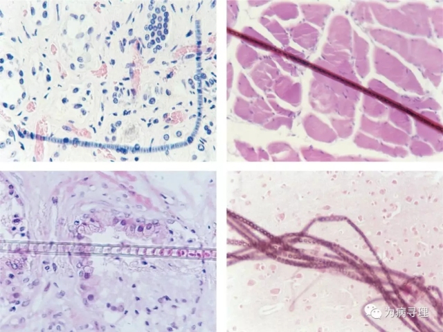
Figure 1A shows the presence, in a kidney histological section, of a filamentous structure formed by a single row of several rectangular/cuboidal blue cells (Σ7 μm in width) arranged end to end. On the basis of the morphology, this structure was catalogued as a member of the Cyanobacteria (blue-green fresh algae), belonging to the genus Oscillatoria, family Oscillatoraceae, class Chroobacteria. | 图1A显示在肾脏组织切片中,由一排首尾相连的单列数个矩形/立方蓝色细胞形成的丝状结构。根据形态特征,该结构被归为蓝藻(蓝绿色新鲜藻类)一员,为Oscillatoria属,Oscillatoraceae科,Chroobacteria纲。 |
Figure 1B shows the presence, in a skeletal muscle histological section, of a non-branched filamentous structure of a blackish hue formed by a chain of cylindrical barrel-shaped cells (4~6μm in length) with a gelatinous sheath of reddish hue. On the basis of the morphology, this structure was catalogued as a member of the Cyanobacteria, belonging to the genus Anabaena, order Nostocales, class Homogoneae. | 图1B显示骨骼肌组织切片中存在一种黑色的非分枝丝状结构,由一系列圆柱形筒状细胞(长4~6μm)形成,鞘呈红色,胶状。在形态学基础上,将其归为蓝藻一员,为Anabaena属,Nostocales目,Homogoneae纲。 |
Figure 1C shows the presence, in a uterine cervix histological section, of a non-branched elongated structure with thick walls, formed by numerous cuboidal cells (7~12μm in width). Each cell possesses one chloroplast located at the margin. Slight narrowings along the side walls can also be observed. On the basis of the morphology, this structure was catalogued as a green fresh alga, belonging to the genus Ulothrix, class Ulvophyceae, division Chlorophyta. | 图1C显示子宫颈组织切片中存在由许多立方细胞(宽7~12μm)形成的壁厚的无分枝细长结构。每个细胞边缘有一个叶绿体。还可以观察到沿侧壁的轻微变窄。根据形态特征,该结构被归为一种绿色的淡水藻类,为Ulothrix属,Ulvophyceae纲, division Chlorophyta。 |
Figure 1D shows the presence, in a brain histological section, of six non-branched filamentous structures, slightly twisted, of a darkish hue, each formed by a row of cuboidal cells (0.8~2μm in length) with clear cytoplasm and with narrow intercellular spaces. The cells are surrounded by a thin gelatinous sheath. On the basis of the morphology, this structure was catalogued as a member of the Cyanobacteria, belonging to the genus Phormidium, family Oscillatoraceae, class Cyanophyceae. | 图1D显示,在脑组织切片中,存在六个微扭曲的暗色调的非分枝丝状结构,每一个结构由一排胞质清晰、细胞间隙狭窄的立方细胞(长0.8~2μm)组成。细胞被一层薄薄的胶状鞘所包围。根据其形态特征,归为蓝藻一员,为Phormidium属,Oscillatoraceae科,Cyanophyceae纲。 |
Similarly, in cytological smears, the presence of diverse types of freshwater algae as contaminants has been reported, the cause also being contamination of tap water. | 同样,在细胞学涂片中,也有不同类型的淡水藻类作为污染物的报道,其原因也是自来水受到污染。 |
Eutrophication is one of the causes of the deterioration of water quality. Thus, the contamination of reservoirs by freshwater algae might explain the presence of these microorganisms in the tap water used not only in laboratories, but also in hospitals as drinking water. Consequently, the significance of this finding is two-fold. | 富营养化是导致水质恶化的原因之一。因此,淡水藻类对水库的污染可能解释了这些微生物在自来水中的存在,这些自来水不仅用作实验室用水,也用作医院的饮用水。因此,这一发现的意义是双重的。 |
Some species of freshwater algae (mainly members of the Cyanobacteria) can produce powerful toxins, such as neurotoxic alkaloids (anatoxins), peptide hepatotoxins (microcystins), and cytotoxic alkaloids (cylindrospermopsins), which present a risk to human health. On the other hand, and because of their morphology, freshwater algae such as those described in this article may be misidentified as filamentous fungi or larvae of certain parasitic worms (e.g. filarial and rhabditiform nematodes). | 一些淡水藻类(主要是蓝藻的成员)能够产生强大的毒素,如神经毒性生物碱(毒伞肽)、肽类肝毒素(microcystins)和细胞毒性生物碱(cylindrospermopsins),这对人体健康构成危险。另一方面,由于其形态,如本文所述的淡水藻类可能被误认为是丝状真菌或某些寄生蠕虫的幼虫。(如丝虫和rhabditiform nematodes) |
As is the case with other types of contaminant (‘floaters’), that may arise during tissue processing and slide preparation and be a potential cause of misdiagnosis in surgical pathology, we think that the presence of freshwater algae in histopathological slides because of eutrophication and their consequent presence in laboratory tap water, should also be of concern to, and recognised by, cellular pathologists. | 与其他类型的污染物(“漂浮物”)一样,在组织处理和玻片制备过程中可能出现,并且可能是外科病理学误诊的潜在原因,我们认为,由于富营养化,组织病理学玻片中存在淡水藻类,以及它们在实验室自来水中的存在,也应该引起细胞病理医生的关注和认可。 |
Figure 1. Histological sections showing the presence of diverse types of freshwater algae. A(左上), Kidney, genus Oscillatoria [haematoxylin and eosin (HE)]. B(右上), Skeletal muscle, Cyanobacteria (HE). C(左下), Uterine cervix, Ulothrix (HE). D(右下), Brain, Phormidium (H&E). | |
参考文献: [1] Eutrophication in histopathology.Histopathology,2019,75,137-138. DOI: 10.1111/his.13852. | |
我要评论


共0条评论