

我的博文
第一集:四大《痛性皮下结节》
(一)血管球瘤 | |
临床 表现 | 好发于20~40岁成人,球血管瘤(glomangioma)多见于儿童。好发于肢体远端的动静脉吻合支,即正常血管球细胞所在处,故最好发部位为手指甲床下(女性多见),也常见于手掌、腕部、前臂和足的皮下或浅表软组织内。消化道以胃最常见,全身其他部位也可见。90%病例为孤立性,10%为多发性(多为儿童患者)。临床表现为发作性疼痛(冷刺激或触摸时引发),从患处向外放射。 |
大体形态 | 肿瘤体积小,多在2cm以下。肿瘤周界清晰,多无包膜,质软,灰红。 |
组织 形态学
| 根据瘤细胞、血管结构和平滑肌组织的不同比例,分为三种类型: ①固有球瘤(glomus proper):占75%,界限清楚,由形态一致的圆形细胞组成,呈片状分布在血管之间或呈环状围绕在血管周围,也可呈血管外皮瘤样排列。瘤细胞呈规则的圆形,胞质淡染透明或淡嗜伊红色,细胞边界清晰,PAS染色更明显。圆形核位于细胞中央,间质可伴玻璃样变或黏液样; ②球血管瘤(glomangioma):界限不清,瘤内血管多为扩张的海绵状血管,血管周围的球细胞簇少而菲薄,血管腔内可有血栓或静脉石; ③球血管肌瘤(glomangiomyoma): 除规则的圆形球细胞外,瘤内还含有平滑肌束,球细胞与平滑肌细胞相互之间有过度现象; 除上述经典形态外,部分病例瘤细胞胞质呈嗜酸性/上皮样,也称嗜酸细胞性血管球瘤(oncocytic glomus tumor)或上皮样血管球瘤(epithelioid glomus tumor); 如瘤细胞的核因退变而具有明显的异型性时,称为共质体性或奇异性血管球瘤(symplastic or bizarre glomus tumor),但无核分裂象或坏死,类似于陈旧性神经鞘瘤或奇异性平滑肌瘤。 如见类似血管外皮瘤样的分支或鹿角状血管,瘤细胞可呈梭形,称为球周皮细胞瘤(glomangiopericytoma); |
免疫 表型
| 瘤细胞表达a-SMA、calponin、vimentin和IV型胶原(鸡爪样); 偶可表达CD34、NGFR和髓鞘相关糖蛋白; 一般不表达desmin(偶尔局灶阳性)、AE1/AE3和S100,偶可表达Syn,但不表达CgA,不要误诊为神经内分泌肿瘤。 |
鉴别 诊断 | ① A cellular, epithelioid glomus tumor may occasionally be confused with a solid variant of nodular hidradenoma. In contrast to a glomus tumor, nodular hidradenoma is positive for keratins, EMA, and carcinoembryonic antigen. ② Unlike glomus tumors, intradermal melanocytic nevus (including the pseudovascular variant) is composed of S-100 protein-positive cells. ③ A carcinoma can be excluded based on clinical findings, reactivity for keratin and EMA, and negativity for smooth muscle actin. ④ Conventional cavernous hemangioma may superficially resemble glomangioma. However, unlike conventional cavernous hemangioma, glomangioma shows a thin rim of glomus cells situated around the vascular spaces. ⑤ Glomus tumors showing a well-developed smooth muscle component or prominent hemangiopericytoma-like features may be confused with myofibroma, infantile myofibromatosis, or myopericytoma. This distinction is of little importance, however, because these entities are related, benign lesions that lie on a morphologic continuum (Box 6.12).
|
分子遗传学 | NOTCH gene rearrangements occur in approximately 60% of glomus tumors. NOTCH2 rearrangements predominate and are identified in most malignant glomus tumors, whereas NOTCH1 and NOTCH3 rearrangements are found in a small subset of predominantly benign glomus tumors. MIR143 has been identified as a fusion partner with NOTCH in some cases. BRAF V600E and KRAS mutations have been reported in a few glomus tumor cases. |
治疗及预后 | 局部切除,切除不净可局部复发。 |
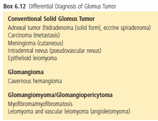
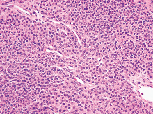
Figure Glomus Tumor. The tumor is composed of rounded cells with sharply defined cell borders.
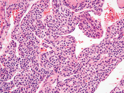
Figure Glomus Tumor. Glomus tumor with prominent branching thin-walled vessels. The tumor cells are situated beneath the endothelium.
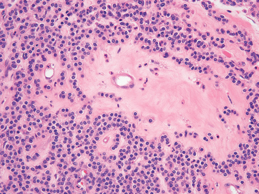
Figure Glomus Tumor. This tumor contains hyalinized stroma. Note the uniform cytology and clear cytoplasm.
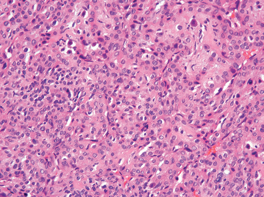
Figure Epithelioid (Oncocytic) Glomus Tumor. The tumor cells contain brightly eosinophilic cytoplasm(Right).
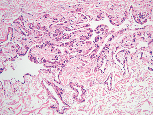
Figure Glomangioma (Glomuvenous Malformation). The lesion is composed of dilated cavernous venous structures surrounded by glomus cells.
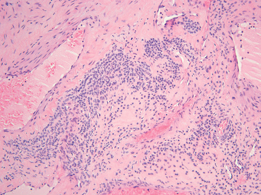
Figure Glomangiomyoma. Glomus cells between thick-walled blood vessels with a prominent smooth muscle component.
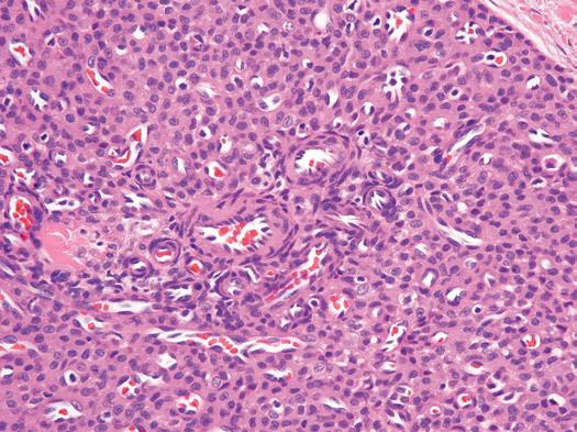
Figure Glomangiopericytoma. In this myopericytoma, many perivascular cells show glomoid morphologic features.
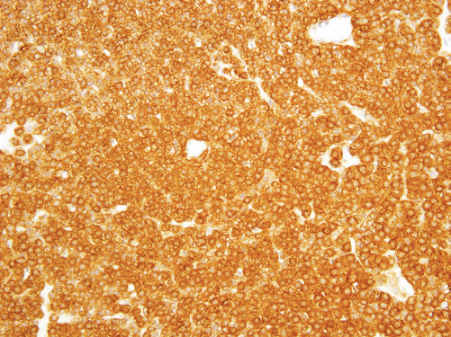
Figure Glomus Tumor. Strong and diffuse expression of smooth muscle actin
by tumor cells.
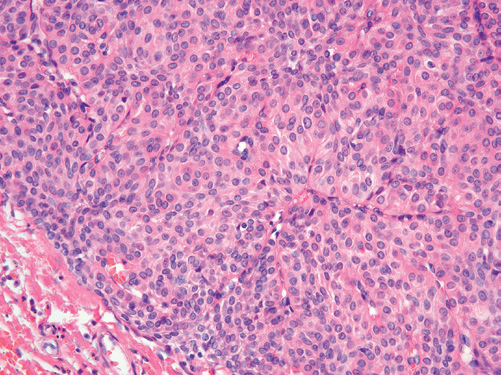
Figure Nodular Hidradenoma. This tumor may be confused with a glomus tumor.
我要评论

共0条评论