我的博文
董俭达:关于胃的乳头状腺癌在不同著作和文献中的描述比较
20170704 银川
2017年7月4日西部消化道早癌导师团空中论坛第二十六期 贵州医科大学附属医院 许良璧主任分享一例胃底多发息肉样病变,问题讨论中对与乳头状腺癌的存在与否提出了问题。本文通过日本胃癌分及WHO胃癌分型的病理学描述回顾对比,为进一步问题的阐述提供资料,共同学习。
一. 来自日本分型。
参考文献:Japanese classification of gastric carcinoma: 3rd English edition
日本胃癌分型第三版2011
2.1.4 Histological classification (Table 4)
Where a malignant epithelial tumor consists of more than one histological subtype, the different histological components should be recorded in descending order of the surface area occupied, e.g., tub 1>pap
组织学分型
恶性上皮来源肿瘤会由多于一种的组织学亚型成分构成,不同的组织学成分在表面区域所占比例进行排序,如表面高分化管状腺癌>乳头状腺癌
Note 2: In clinicopathological or epidemiological studies,papillary or tubular adenocarcinoma can be interpreted as differentiated or intestinal type,whereas "por" and "sig" can be regarded as the undifferentiated or diffuse type. Mucinous carcinoma can be interpreted as either intestinal or diffuse, depending upon the other predominant elements (pap, tub, por or sig).
注意:在临床病理学及流行病学研究中,乳头状或是管状腺癌被定义为分化型的癌或是肠型腺癌。但是低分化腺癌和印戒细胞癌可以被认为是未分化或是弥漫型腺癌。粘液腺癌也可以被分为肠型(分化型)或弥漫型,取决于其主要成分类型(乳头,管状,低分化。。。)
Table 4 Histological classification of gastric tumors
Benign epithelial tumor ICD-O code
Adenoma 8140/0
Malignant epithelial tumor
Common type
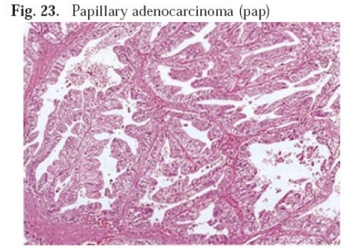
Papillary adenocarcinoma (pap) 8260/3乳头状癌
Tubular adenocarcinoma (tub) 8211/3管状腺癌
Well-differentiated (tub1)高分化
Moderately differentiated (tub2)中分化
Poorly differentiated adenocarcinoma (por)低分化腺癌
Solid type (por1) 实体状
Non-solid type (por2)
Signet-ring cell carcinoma (sig) 8490/3 印戒细胞癌
Mucinous adenocarcinoma (muc) 8489粘液腺癌/3
Special type 特殊类型
Carcinoid tumor 8240/3 类癌
Endocrine carcinoma 8401/3 神经内分泌癌
Carcinoma with lymphoid stroma 髓样癌
Hepatoid adenocarcinoma 肝样腺癌
Adenosquamous carcinoma 8560/3 腺鳞癌
Squamous cell carcinoma 8070/3鳞癌
Undifferentiated carcinoma 8020/3未分化癌
Miscellaneous carcinoma

比较上下两图,在一个管子里,向管子腔内长出了形状不一的乳头结构,绿线是纤维血管轴心,蓝色轮廓是乳头被覆的上皮(肿瘤细胞),还有箭头所指是另一个腺腔内的乳头结构。

一下这个图中,这个管状的可就真的是管状了啦。中间的空白是管腔,外周是上皮细胞(肿瘤)管壁。


上面这个图中分化,管状结构为主,但已经有了筛状和实体状的趋势的区域
20170704 早癌空中论坛许老师一例相关图片:


绿线为正常粘膜,黑圈为肿瘤。右下角一小块组织人工现象。


镜下组织学类型和以上文献比较可以看出是日本分型tub1+tub2,偶见pap结构。
二、来自阿克曼病理学第九版

书中未特别的描述乳头状腺癌。
三、来自WHO-Pathology and genetics of tumours of the digestive system2000
消化系统肿瘤病理及遗传学2000
与WHO2010年版比较没有大的一下内容没有大的改动,新版胃腺癌的WHO分型不再强调肠型和弥漫型这一分型方法,在其他分类体系中简要介绍了Lauren分型(肠型和弥漫型)和其他分型。新版中胃腺癌主要分为乳头状、管状、黏液、黏附性差的癌(包括印戒细胞癌和其他变型)和混合性腺癌。

Histopathology
Gastric adenocarcinomas are either gland-forming malignancies composed of tubular, acinar or papillary structures, or they consist of a complex mixture of discohesive, isolated cells with variable morphologies, sometimes in combination with glandular, trabecular or alveolar solid structures
组织学
胃腺癌既包括管状、腺泡状及乳头状的构成的恶性腺样结构,还包括好一些混合性的失粘附性的游离多形性细胞的复杂结构,有时同时包含有腺样、管状及泡状实体样结构。
Papillary adenocarcinomas
These are well-differentiated exophytic carcinomas with elongated finger-like processes lined by cylindrical or cuboidal cells supported by fibrovascular connective tissue cores. The cells tend to maintain their polarity. Some tumours show tubular differentiation (papillotubular). Rarely, a micropapillary architecture is present. The degree of cellular atypia and mitotic index vary; there may be severe nuclear atypia. The invading tumour edge is usually sharply demarcated from surrounding structures; the tumour may be infiltrated by acute and chronic inflammatory cells.
乳头状腺癌
指的是那些高分化的外生性生长的癌伴有伸长的指状突起,其表面被覆有柱状细胞并由纤维血管轴心支持营养的结构。细胞保持了极性排列(方向一致)。有的肿瘤表现为管状分化(乳头状管状)。少见情况是,微乳头状结构的出现。细胞异型性和增殖指数低,也可见高度异性型细胞。肿瘤与周围组织边界清,肿瘤内可以看到急性及慢性炎症细胞浸润。
通过以上的介绍, “20170704 早癌空中论坛许老师一例”1、局部看到肿瘤表面较为平坦,未见明显的指状突起,无论细长或是粗大乳头结构,仅见平坦裂隙样分开的腺样结构,这时低倍下的外生性生长的结构也可以认为非常粗大的外生乳头样结构,这样的意义如何,可能和内镜下所见去对应观察会更有意义。2、本例中“腺管内的乳头状分化”的结构不是很典型(日本分型中的乳头状腺癌演示图)。3、微乳头结构也没有看到(实际上微乳头结构形成常常与肿瘤的高侵袭力及更厉害的生物学行为有关)。所以,本例目前资料来看不是,最起码可以说不是典型的乳头状腺癌,这样外生性生长模式,可以和肠道肿瘤很相似,出现绒毛状、指状、花菜样、蘑菇样。。。。。乳头状等等的外生性生长特征,及腺管内的乳头状生长,这些可能更多都是腺癌高分化或肠型腺癌的特征。出现微乳头结构或是混杂有低分化癌、印戒细胞癌等成分是需要进一步评估和描述的。
仓促准备,请各位老师批评指正。
我要评论


共0条评论